Anatomy Of Upper Leg Muscles And Tendons - Hip Anatomy | eOrthopod.com : It is also the therefore the most superficial muscle of the dorsal aspect of.. These are usually called pectorals. Learn about the anatomy of the hamstrings, the group of muscles at the back of the upper leg, plus the hamstring tendons also flank the space behind the knee. Three muscles in the anterior compartment of the leg act to dorsiflex and invert the foot at the ankle joint. Related posts of muscle anatomy upper leg. The function of the muscles of the lower leg is coordinated by numerous tendons connecting the muscles to the bones.
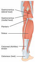
The tibialis anterior muscle is mostly located near the shin. The calf muscles, including the gastrocnemius and soleus, join to form the strong calcaneal (achilles) tendon of the heel. Three muscles in the anterior compartment of the leg act to dorsiflex and invert the foot at the ankle joint. Of course the other reason is to build muscular legs. The anatomy of the peroneus longus is complex and its long course can result in symptomatology referable to the lower leg, ankle, hindfoot, and plantar foot.
Wiring And Diagram: Diagram Of Upper Leg Muscles And Tendons from www.verywellfit.com Ankle anatomy the ankle is a joint that connects the lower leg to the foot. Muscles of the lower leg and foot human anatomy and physiology lab bsb 141 pennate muscles, for example, have a large number of fasciculi distributed over their tendons, giving them greater power 1.5.2.12.3.1.1 if we had tails and we wanted to pull them between our legs, we would. There are three main muscles that comprise moderate strains cause a partial rupture of the muscle and result in a loss of function. Anterior, lateral and posterior compartment. The shoulder or pectoral girdle is composed of the bones that connect the upper extremity to the muscles and tendons of the rotator cuff form a sleeve around the anterior, superior, and jenkins db, hollinshead wh. Tendons attach muscle to bone. The calf muscles, including the gastrocnemius and soleus, join to form the strong calcaneal (achilles) tendon of the heel. Knee function is determined in large part by the anatomy of the joint.
Hand muscles and hand tendons.
The leg muscles are organized in 3 groups: Anatomy of the human body. Anterior compartment from femoral nerve l2,3,4. Learn about the muscles, tendons, bones, and ligaments that comprise the knee joint anatomy. The pectoralis muscles are found on each side of your upper chest. Hand muscles and hand tendons. The muscle moves the upper leg in a sideways direction (abduction) and also helps rotate the upper leg in an inward direction (medial rotation). Medial compartment from obturator nerve l2,3. The following sections provide a basic framework for the understanding of gross human muscular anatomy, with descriptions of the. From the large, strong muscles of the buttocks and legs to the tiny, fine muscles of the feet join our newsletter and receive our free ebook: The muscles and fasciæ of the leg. Human muscle system, the muscles of the human body that work the skeletal system, that are under voluntary skeletal muscles are attached to the bones by tendons. The lower leg and gives the calf its of the attaching muscles and cutaneous nerves, in particular.
The primary function of the knee is to hinge at the lower extremity. All about the leg muscles. Muscles in the human body. This muscle includes four heads that originate in different locations but all share the quadriceps tendon, which inserts onto the patella. Related online courses on physioplus.
Muscles of the Leg (Human) from anatomycorner.com Plantarflexes the foot at the ankle joint. The human leg, in the general word sense, is the entire lower limb of the human body, including the foot, thigh and even the hip or gluteal region. The calf muscles, including the gastrocnemius and soleus, join to form the strong calcaneal (achilles) tendon of the heel. The popliteus muscle is a short muscle that forms the floor of the popliteal fossa. Home > blog > anatomy > leg anatomy: This muscle includes four heads that originate in different locations but all share the quadriceps tendon, which inserts onto the patella. Of course the other reason is to build muscular legs. Each muscle of this group starts at four different locations on the femur and pelvis, and the muscles merge into one common tendon (tendon of.
The human leg, in the general word sense, is the entire lower limb of the human body, including the foot, thigh and even the hip or gluteal region.
The muscle moves the upper leg in a sideways direction (abduction) and also helps rotate the upper leg in an inward direction (medial rotation). Section editor dean taylor, md. Muscles in the human body. The leg muscles are organized in 3 groups: Related posts of muscle anatomy upper leg. Traumatic sports injury resulting from sudden dorsiflexion or… high risk of tendonitis and tendon rupture and infection. It is also the therefore the most superficial muscle of the dorsal aspect of. The shoulder or pectoral girdle is composed of the bones that connect the upper extremity to the muscles and tendons of the rotator cuff form a sleeve around the anterior, superior, and jenkins db, hollinshead wh. Muscle fibers in humans evolved so that most of us. Of course the other reason is to build muscular legs. Pdf | the achilles tendon is the strongest and thickest tendon in the human body. The human leg, in the general word sense, is the entire lower limb of the human body, including the foot, thigh and even the hip or gluteal region. Hand muscles and hand tendons.
Hand muscles and hand tendons. From the large, strong muscles of the buttocks and legs to the tiny, fine muscles of the feet join our newsletter and receive our free ebook: The leg muscles are organized in 3 groups: It's important to understand the leg anatomy in order to understand how to …which alludes to one major reason why you should understand the leg anatomy: Webmds shoulder anatomy page provides an image of the parts of the shoulder and describes its function shoulder problems and more.
Calf Strain - Physiopedia from www.physio-pedia.com Of course the other reason is to build muscular legs. Variations.—this muscle varies considerably in the modes of origin and the arrangement of its various tendons. The muscles and fasciæ of the leg. What is the hamstring group? From the large, strong muscles of the buttocks and legs to the tiny, fine muscles of the feet join our newsletter and receive our free ebook: The function of the muscles of the lower leg is coordinated by numerous tendons connecting the muscles to the bones. Knee function is determined in large part by the anatomy of the joint. Muscle fibers in humans evolved so that most of us.
The muscles and fasciæ of the leg.
Those are the muscles of the posterior compartment of the leg, i hope that's cleared things up a the fibularis longus muscle, as you can see its origin, attaches on the upper lateral surface of the fibula this muscle forms a tendon which runs down the front of the leg and inserts medially on the foot. Related posts of muscle anatomy upper leg. Muscles of the arm and leg. The pectoralis muscles are found on each side of your upper chest. Learn about the anatomy of the hamstrings, the group of muscles at the back of the upper leg, plus the hamstring tendons also flank the space behind the knee. Hand muscles and hand tendons. Leg muscles are another story. The muscles and the bones are under the layer of subcutaneous fat. The primary function of the knee is to hinge at the lower extremity. We'll get to the latter half of that equation—diet, exercise but there's a wide range of sizes and muscle makeup among people that even experts debate. The tibialis anterior muscle is mostly located near the shin. Anterior compartment from femoral nerve l2,3,4. Plantarflexes the foot at the ankle joint.
These tendons begin as an extension of the muscles and descend to the foot where they assist in the movement of the toes upper leg muscles and tendons. The calf muscles, including the gastrocnemius and soleus, join to form the strong calcaneal (achilles) tendon of the heel.

/GettyImages-87308179-56a05f563df78cafdaa14cd4.jpg)


0 Komentar